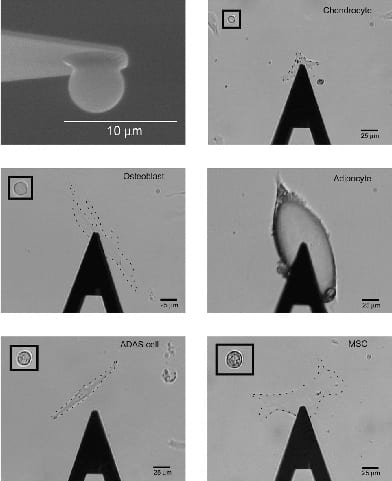
The mechanical characteristics associated with individual cells can reveal much about what they are and how they function in an organism. While many researchers are interested in how mechanical forces affect cells or how the forces cells exert affect their surrounding environment, the Darling Lab focuses on the intrinsic mechanical properties exhibited by specific cell types in unperturbed and perturbed situations. This “mechanophenotype” serves as a biomarker of sorts and is a secondary indicator of many complex, biological events occurring inside the cell. We have investigated the mechanophenotype of many cell types (1-9), and how these characteristics change in response to environmental stimuli (10-14). We are also studying how “mechanical biomarkers” can be applied to cell sorting/enrichment (15-17). Single-cell elastic and viscoelastic properties are obtained using atomic force microscopy (AFM), which allows for high-resolution force and displacement measurement at extremely small scales. We also use AFM to examine the microscale mechanical properties of cell clusters and engineered or isolated tissues (18-20) to complement a range of biochemical assays to answer hypothesis-driven studies.
- Dempsey ME, Chickering GR, González-Cruz RD, Fonseca VC, Darling EM. (2022) Discovery of surface biomarkers for cell mechanophenotype via an intracellular protein-based enrichment strategy. Cell Mol Life Sci. 79(320). PMCID: Pending. DOI: 10.1007/s00018-022-04351-w
- Gutierrez RA, Fang W, Kesari H*, Darling EM*. (2021) Force sensors for measuring microenvironmental forces during mesenchymal condensation. Biomaterials. 270: 120684. PMCID: PMC7906959. DOI: 10.1016/j.biomaterials.2021.120684 (*Co-corresponding author)
- Dubay R, Fiering J, Darling EM. (2020) Effect of elastic modulus on inertial displacement of cell-like particles in microchannels. Biomicrofluidics. 14(4): 044110. PMCID: PMC7402708. DOI: 10.1063/5.0017770
- González-Cruz RD, Dahl KN, Darling EM. (2018) The Emerging Role of Lamin C as an Important LMNA Isoform in Mechanophenotype. Frontiers Cell Devel Biol. 6: 151. PMCID: PMC6224339. DOI: 10.3389/fcell.2018.00151
- Labriola NR, Sadick JS, Morgan JR, Mathiowitz E, Darling EM. (2018) Cell mimicking microparticles influence the organization, growth, and mechanophenotype of stem cell spheroids. Ann Biomed Eng. 46(8): 1146-1159. PMCID: PMC6039261. DOI: 10.1007/s10439-018-2028-4
- González-Cruz RD, Sadick JS, Fonseca VC, Darling EM. (2018) Nuclear lamin protein C is linked to lineage-specific, whole-cell mechanical properties. Cell Mol Bioeng. 11(2): 131-142. PMCID: PMC5943047. DOI: 10.1007/s12195-018-0518-y
- Shah MK, Garcia-Pak IH, Darling EM. (2017) Influence of inherent mechanophenotype on competitive cellular adherence. Ann Biomed Eng. 45(8): 2036-2047. PMCID: PMC5529242. DOI: 10.1007/s10439-017-1841-5
- Yang X, Darling EM, Herzog W. (2017) Functional properties of chondrocytes and articular cartilage using optical imaging to ccanning probe microscopy. J Orthop Res. PMCID: PMC5839958. DOI: 10.1002/jor.23757
- López-Fagundo C, Livi LL, Ramchal T, Darling EM*, Hoffman-Kim D*. (2016) A biomimetic synthetic feeder layer supports the proliferation and self-renewal of mouse embryonic stem cells. Acta Biomater. 39: 55-64. PMCID: PMC4905775. DOI: 10.1016/j.actbio.2016.04.047 (*Co-corresponding author)
- Chen Y, Cossman J, Jayasuriya CT, Li X, Guan Y, Fonseca VC, Yang K, Charbonneau C, Yu H, Kanbe K, Ma P, Darling EM, Chen Q. (2016) Deficient mechanical activation of anabolic transcripts and post-traumatic cartilage degeneration in matrilin-1 knockout mice. PLoS ONE. 11(6): E0156676. PMCID: PMC4896629. DOI: 10.1371/journal.pone.0156676
- Dingle YL, Chirila AM, Boutin ME, Livi LL, Labriola NR, Jakubek LM, Morgan JR, Darling EM, Kauer JA, Hoffman-Kim D. (2015) Three-Dimensional Neural Spheroid Culture: An In Vitro Model for Cortical Studies. Tissue Eng C. 21 (12): 1274-83. PMCID: PMC4663656. DOI: 10.1089/ten.tec.2015.0135
- Darling EM, DiCarlo D. 2015. High-throughput assessment of cellular mechanical properties. Ann Rev Biomed Eng. 17:35-62. PMCID: PMC8204286. DOI: 10.1146/annurev-bioeng-071114-040545
- Labriola NR, Darling EM. 2015. Temporal heterogeneity in single-cell gene expression and mechanical properties during adipogenic differentiation. J Biomech. 28(2015): 1058-66. PMCID: PMC4380682. DOI: 10.1016/j.jbiomech.2015.01.033
- Kanthilal M, Darling EM. (2014). Characterization of mechanical and regenerative properties of human, adipose stromal cells. Cell Mol Bioeng. 7 (4): 585-97. PMCID: PMC4255916. DOI: 10.1007/s12195-014-0350-y
- Toyjanova J, Hannen E, Bar-Kochba E, Darling EM, Henann D, Franck C. (2014). 3D viscoelastic traction force microscopy. Soft Matter. 10: 8095-106. PMCID: PMC4176508. DOI: 10.1039/C4SM01271B
- González-Cruz RD, Darling EM. (2013). Adipose-derived stem cell fate is predicted by cellular mechanical properties. Adipocyte. 2 (2): 87-91. PMCID: PMC3661107. DOI: 10.4161/adip.23015
- González-Cruz RD, Fonseca VC, Darling EM. (2012). Cellular mechanical properties reflect differentiation potential of adipose-derived mesenchymal stem cells. Proc Natl Acad Sci USA. 109 (24): E1523-9. PMCID: PMC3386052. DOI: 10.1073/pnas.1120349109
- Darling EM. (2011). Force scanning: A rapid, high-resolution approach for spatial mechanical property mapping. Nanotechnology. 22 (17) 175707. PMCID: PMC3150532. DOI: 10.1088/0957-4484/22/17/175707 *Recognized by Nanotechnology in their “Highlights 2011” Collection.
- Gilchrist CL, Darling EM, Chen J, Setton LA. (2011). Extracellular matrix ligand and stiffness modulate immature nucleus pulposus cell‐cell interactions. PLoS One. 6 (11): e27170. PMCID: PMC3210142. DOI: 10.1371/journal.pone.0027170
- Yim EKF, Darling EM, Kulangara K, Guilak F, Leong KW. (2010). Nanotopography-induced changes in focal adhesions, cytoskeletal organization, and mechanical properties of human mesenchymal stem cells. Biomaterials. 31 (2010): 1299-1306. PMCID: PMC2813896. DOI: 10.1016/j.biomaterials.2009.10.037
- Darling EM*, Wilusz RE*, Bolognesi MP, Zauscher S, Guilak F. (2010). Spatial mapping of the biomechanical properties of the pericellular matrix of articular cartilage measured in situ via atomic force microscopy. Biophys J. 98: 2848-2856. PMCID: PMC2884253. DOI: 10.1016/j.bpj.2010.03.037 (*co-first author)
- Darling EM, Pritchett PE, Evans BA, Superfine R, Zauscher S, Guilak F. (2009). Mechanical properties and gene expression of chondrocytes on micropatterned substrates following dedifferentiation in monolayer. Cell Molec Bioeng. 2 (3): 395-404. PMCID: PMC2898162. DOI: 10.1007/s12195-009-0077-3
- Coles J, Blum J, Jay G, Darling EM, Guilak F, Zauscher S. (2008) In situ friction measurement on murine cartilage by atomic force microscopy. J Biomech. 41 (3): 541-548. PMCID: PMC2274896. DOI: 10.1016/j.jbiomech.2007.10.013
- Darling EM, Topel M, Zauscher S, Vail TP, Guilak F. (2008) Viscoelastic properties of human mesenchymally-derived stem cells and primary osteoblasts, chondrocytes, and adipocytes. J Biomech. 41 (2): 454-464. PMCID: PMC2897251. DOI: 10.1016/j.jbiomech.2007.06.019
- Darling EM, Zauscher S, Block JA, Guilak F. (2007) A thin-layer model for viscoelastic, stress-relaxation testing of cells using atomic force microscopy: Do cell properties reflect metastatic potential? Biophys J. 92: 1784-1791. PMCID: PMC1796808. DOI: 10.1529/biophysj.106.083097
- Darling EM, Zauscher S, Guilak F. (2006) Viscoelastic properties of zonal articular chondrocytes measured by atomic force microscopy. Osteoarthritis Cartilage. 14 (6): 571-579. PMID: 16478668. DOI: 10.1016/j.joca.2005.12.003