RAPID VISUALIZATION ASSAY

Methodology

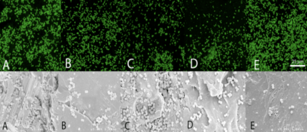
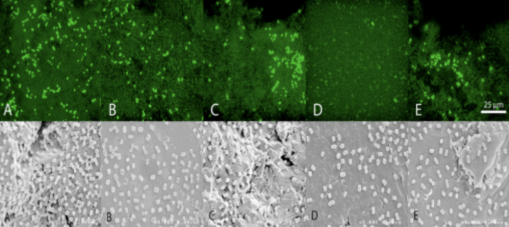
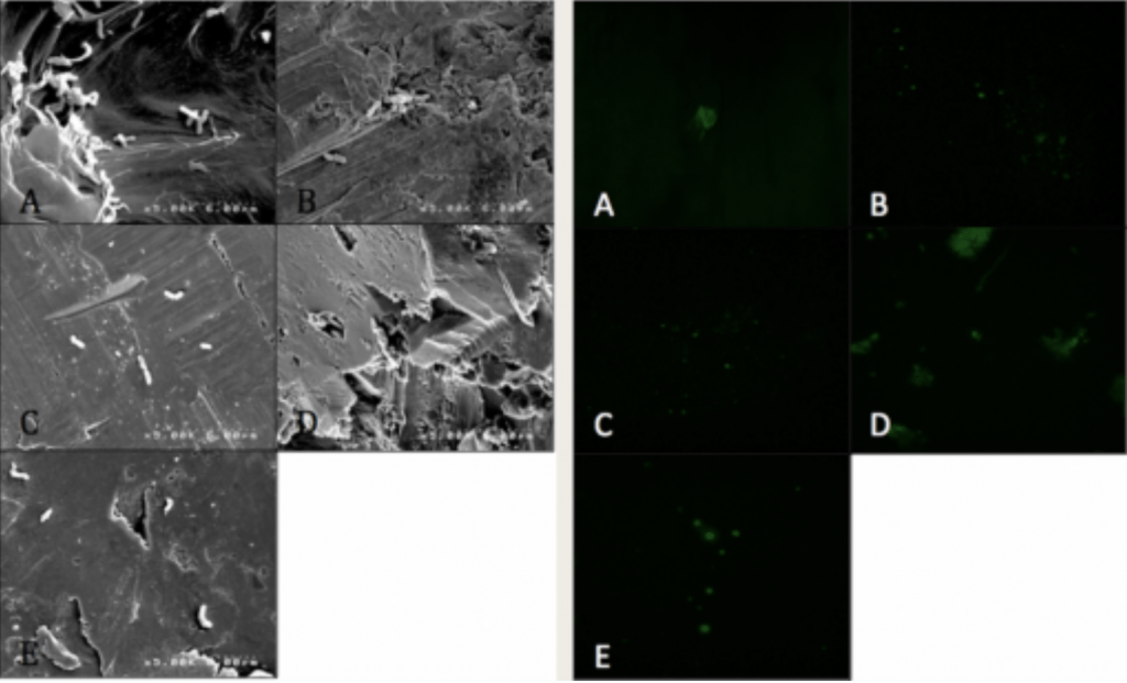
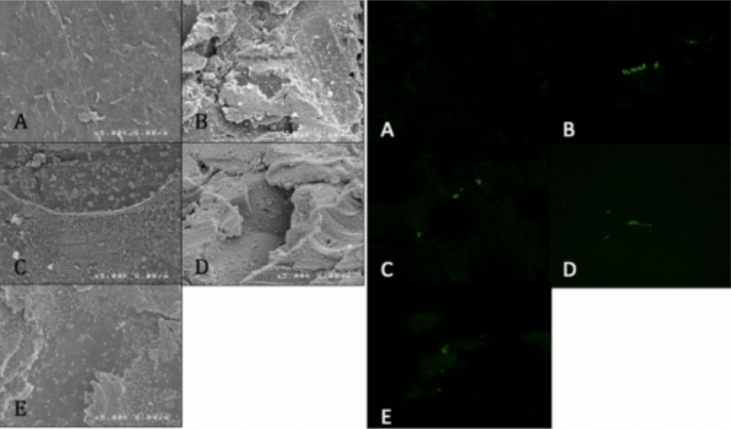
  • Confocal Laser-Scanning microscopy and conjugated fluorescent antibodies.
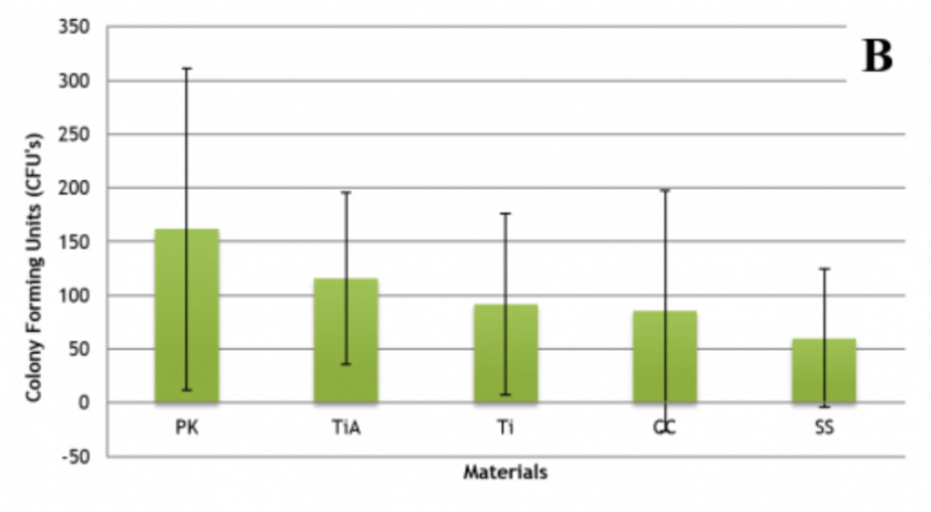
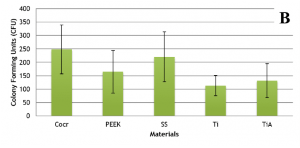
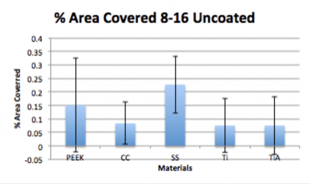
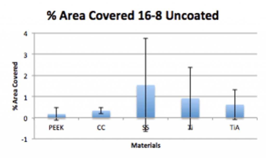
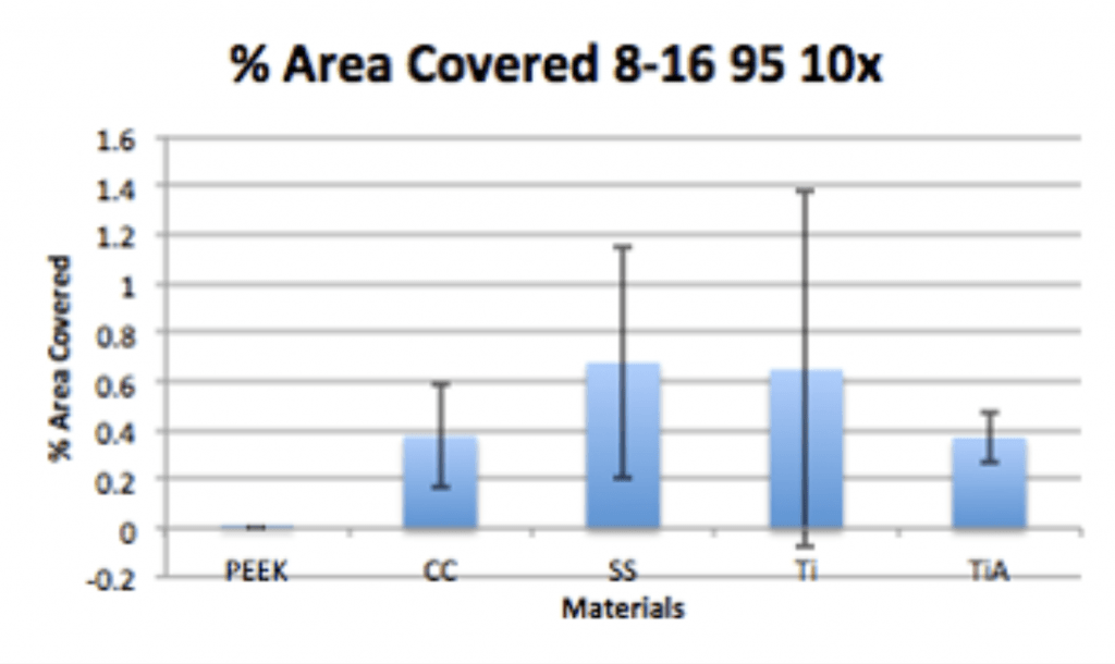
    • Visualize and quantify microbial adherence directly on orthopaedic explants.
    • Optical Sectioning of Topographically Complex Samples
    • Electronic Reconstruction


Design of FCab and Confocal Microscopy
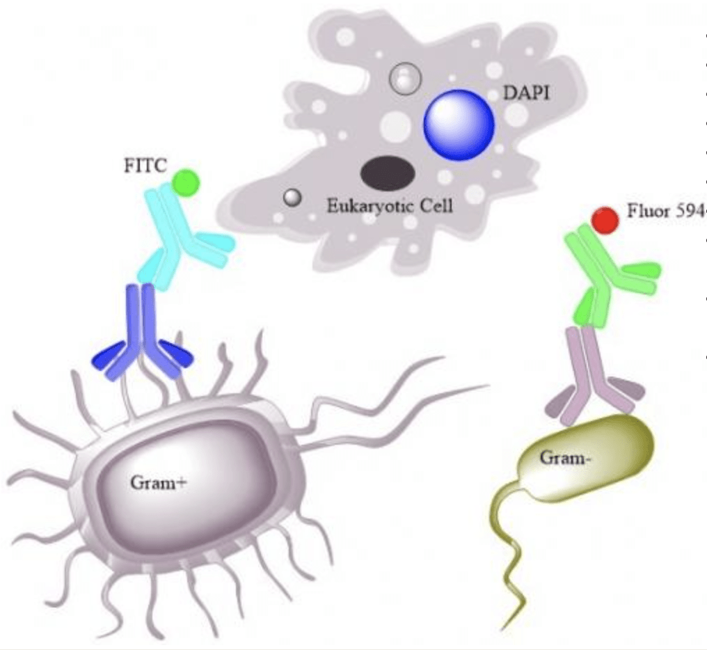
Antibody Tagging
  • Gram Positive
  • Fluorescein Isothiocyanate (FITC)
  • Ex 492nm-Em 518nm (Green)
  • Gram Negative
  • Alexa Fluor 594
  • Ex 590nm-Em 617nm (Red)
  • Eukaryotic Tissue
  • 4’, 6-Diamidino-2-Phenindole, Dihydrochloride (DAPI)
  • Binds to AT-rich regions of DNA (Nuclear stain)
  • Ex 358nm-Em 461nm (Blue)

BACTERIAL ADHERENCE AND BIOFILM STUDY

Characterization of the Adherence Patterns and Biofilm Density of Commonly Encountered Bacterial Pathogens to Spinal Instrumentation of Differing Compositions

Background

  • Infections are one of the most pressing problems in orthopaedic surgery.
  • Very little information on ability of implant materials to minimize infection.
  • Grow most prevalent orthopaedic pathogens on the most commonly utilized orthopaedic implant materials.
  • Utilize Scanning Electron Microscopy (SEM) and Confocal-Laser Scanning Microscopy (CLSM) with fluorescent-conjugated antibodies to determine adherence density and patterns on materials.

Methodology

  • 6 Clinically-Isolated Pathogens
    • Methicillin-Resistant S. aureus
    • Methicillin-Sensitive S. aureus
    • Coagulase-Negative S. epidermidis
    • Multidrug-Resistant A. baumannii
    • Propionibacterium acnes
    • Vancomycin-Resistant E. faecalis 
  • 5 Commonly Utilized Spinal Implant Materials
    • Polyetheretherketone (PEEK)
    • Cobalt Chromium (CoCr)
    • Stainless Steel (SS)
    • Titanium (Ti)
    • Titanium Alloy (TiA)
  • Microscopy Techniques
    • Scanning Electron Microscopy  (SEM)
    • Confocal Laser Scanning Microscopy (CLSM) and Fluorescent-Conjugated Antibodies (FCAs)

Results

Fig 1.  Area  coverage and density of colony forming units (CFUs) of Methicillin-Resistant S. aureus  A) Titanium B) Stainless Steel C) Cobalt Chromium D) PEEK E) Titanium Alloy.

Fig 2 A. % Area Coverage of  Methicillin-Resistant S. aureus.

Fig 2 B. CFU per area of Methicillin-Resistant S. aureus.

Fig 3. Area  coverage and density of colony forming units (CFUs) of Multi-Drug Resistant A. baumannii A) Titanium B) Stainless Steel C) Cobalt Chromium D) PEEK E) Titanium Alloy.

Fig 4. A. % Area Coverage of  Multi-Drug Resistant A. baumannii.

Fig 4 B. CFU per area of Multi-Drug Resistant A. baumannii.
Fig 5. Area  coverage and density of colony forming units (CFUs) of Coagulase-Negative S. epidermidis A) Titanium B) Stainless Steel C) Cobalt Chromium D) PEEK E) Titanium Alloy.

Fig 6 A. % Area  coverage of Coagulase-Negative S. epidermidis.

Fig 6 B. CFU per area of Coagulase-Negative S. epidermidis.

Conclusion

  • No single implant material is able to reduce bacterial adherence of all of the pathogens tested.
  • Cobalt Chromium and PEEK have highest level of bacterial adherence.
  • Stainless Steel and Titanium Alloy have the least amount of bacterial adherence.
  • Adherence appears to be guided by surface microtopography of materials.

DETERMINATION OF ADHERENCE KINETICS OF P. ACNES AND EFFICACY OF TIO2-PDMS SILVER COATING

Background

Propionibacterium acnes

  • Slow-growing, anaerobic-aerotolerant, gram positive rod.
  • Commonly found as part of the natural flora of the large intestine, conjunctiva, oral cavity, and in the pilosebaceous follicles of the skin.
    • Most common organism colonizing shoulder area.
  • Commonly recognized as the causative agent of acne vulgaris.
  • Recognized as an opportunistic pathogen colonizing implants on the shoulder region.
    • Most frequently isolated pathogen in prosthetic shoulder joint infections.
  • Current pre-surgical dressings like chlorheximide gluconate appear to not effective against it.

Methodology

  • Cultured in a humidified anaerobic environment in Reduced Clostridial Media (BD) for 48hrs at 37oC.
  • Implants inoculated with 1×10⁷ cfu/ml for variable times of adherence and proliferation.
  • Dehydrated, Fixed, and Labeled.
  • Visualized via SEM and CLSM.
  • Coated implants dip-coated in 95% 10X (95% TiO2: 5% PDMS) and 100% Ag.

Experimental Conditions

4-20 8-16 12-12 16-8 20-4
Table 1.  Adherence and Proliferation times for PA.  The first number in each set corresponds to the number of hours allowed for adherence, while the second number corresponds to the number of hours allowed for proliferation. Each of these conditions was examined for each coating condition tested.
Control Experimental Control
Uncoated 95% 10X 100% Ag
Table 2. Coating conditions for PA experiments. Concentration ratios of TiO2 to PDMS doped with silver. 95% refers to TiO2 to PDMS ratio, while 0x and 10x correspond to silver neodecanoate concentrations. 100% Ag refers to coating with no TiO2/PDMS. 

Results

Spinal Implant Materials

Fig 1.  100x magnification of implant materials after machining, non-abrasive cleaning, and autoclave sterilization. (A) Ti, (B) SS, (C) CC, (D) PEEK, (E) TiA.

Fig 2. Contact Angle of Spinal Implant Materials


Fig 3. Representative SEM images at 5,000x magnification (Left) and Confocal Laser Scanning Microscopy at 120x (right) of P. acnes on native material surfaces.  (A) PEEK, (B) CC, (C) SS, (4) Ti, (5) TiA. (4-20)

Fig 4. Representative SEM images at 5,000x magnification (Left) and Confocal Laser Scanning Microscopy at 120x (right) of P. acnes on native material surfaces.  (A) PEEK, (B) CC, (C) SS, (4) Ti, (5) TiA. (8-16)


Fig 5. Average number of bacteria on SEM images of uncoated materials at 8 hours adherence and 16 hours proliferation.

Fig 6. Percentage area coverage on confocal microscopy of uncoated materials at 8 hours adherence and 16 hours proliferation.


Fig 7. Representative SEM images at 5,000x magnification (Left) and Confocal Laser Scanning Microscopy at 120x (right) of P. acnes on native material surfaces.  (A) PEEK, (B) CC, (C) SS, (4) Ti, (5) TiA. (16-8)

Fig 8. Average number of bacteria on SEM images of uncoated materials at 16 hours adherence and 8 hours proliferation.

Fig 9. Percentage area coverage on confocal microscopy of uncoated materials at 16 hours adherence and 8 hours proliferation.

Fig 10. Representative SEM images at 5,000x magnification (Left) and CLSM at 120x (Right) of P. acnes on 100% Ag coated surfaces.  (A) PEEK, (B) CC, (C) SS, (4) Ti, (5) TiA (8-16).

Fig 11. Average number of bacteria on SEM images of 100% Ag coated materials at 8 hours adherence and 16 hours proliferation.

Fig 12. Percentage area coverage on confocal microscopy of 100% Ag coated materials at 8 hours adherence and 16 hours proliferation.

Fig 13. Representative SEM images at 5,000x magnification (Left) and CLSM at 120x (Right)

Fig 14. Average number of bacteria on SEM images of 95 10x coated materials at 8 hours adherence and 16 hours proliferation.

Fig 15. Percentage area coverage on confocal microscopy of 95 10x coated materials at 8 hours adherence and 16 hours proliferation.

Fig 16. Representative samples of uncoated pigskin after H&E staining and histopathology at 20x magnification.

Fig 17. Representative samples of pigskin coated in chlorhexidine gluconate after H&E staining and histopathology at 20x magnification.
Fig 18. Representative samples of pigskin coated in silver doped TiO2-PDMS after H&E staining and histopathology at 20x magnification.

Conclusion

  • P. acnes is able to form a biofilm within 4hrs of adherence and 20hrs of propagation on PEEK implants.
  • Ag-Doped Coating is able to prevent biofilm formation and reduce overall bacterial adhesion.
  • Ag-Doped Coating can penetrate deeper into pilosebaceous follicles than chlorheximide gluconate.

ASSESSING THE EFFICACY OF A SILVER-DOPED ANTIMICROBIAL COATINGS ON PROSTHETIC LINERS

Background

Problems with Day to Day Prosthetic Liner Wear

  • In the United States, roughly one in every 180 Americans is currently living with a lost limb after amputation.1
  • There are currently two million patients in the United States living with amputation, and with over 130,000 new amputations each year, this number is estimated to nearly double in the next 35 years as projections call for a total amputee population of 3.9 million in the United States by 2050.1
  • Unfortunately, much of the focus for a surgeon while performing amputation is on the short-term outcome of the patient. Currently, a successful amputation means a complication-free procedure which leaves the patient with a properly sealed limb-end fit for prosthesis attachment.2
  • Due to the ease of this bacterial growth in the prosthetic socket, amputation is often coupled with long term bacteria-related complications, most notably skin irritation, ulceration, infection, sepsis, or in extreme cases, death.3
    • Chronic malodor.

Hypothesized Advantages of Coating on Prosthetic Liners

  • Titanium dioxide has been demonstrated to have anti-inflammatory properties.
  • Polydimethylsiloxane (PDMS) confers flexibility and strong binding adherence to soft liners.
  • When dried, coating is approximately 100nm thick.
  • Sweat and humidity generated in the enclosed stump-liner interface will drive silver release.

Methodology

Liners Pathogens
– Alpha Hybrid
– Ottobock Thermoplastic TPE
– Proteor Keasy
– Alpha Silicone
– MRSA
– P. acnes
– VR E. faecalis
– MSSA
– CoNS epidermidis
– MDR A. baumannii
  • 8mm plugs of liners excised with biopsy punch.
  • Plugs stored in 70% ethanol overnight.
  • Dip coated in each respective condition and allowed to cure in the hood overnight.
  • Plates inoculated with a lawn of bacteria at 2×10⁷ CFU/ml.
  • Plugs placed on plate (liner side down) for 72hrs.

Results

Fig 1. Liner surface interaction with coating

Fig 2. A. baumannii dose response curve

Fig 3. Kirby Bauer Assay showing inhibition at the 10X and 100% Ag-doped liner plug

Keasy Kirby Bauer A. baumannii

Thermoplastic Kirby Bauer A. baumannii

Silicone Kirby Bauer A. baumannii

Hybrid Kirby Bauer A. baumannii


Welcome to the Weiss Lab!

About Us:

Our work in the Diane N. Weiss Center for Orthopaedic Trauma Research focuses on providing translational technologies aimed at preventing, treating, and minimizing the problems of infection related to orthopaedic trauma. Our approach emphasizes translational outcomes based on immediate clinical needs. Our general aim is focused on investigating the permitting factors relating to bacterial adherence and biofilm formation on clinically relevant surfaces, as well as means to more efficiently diagnose, treat, and prevent them.