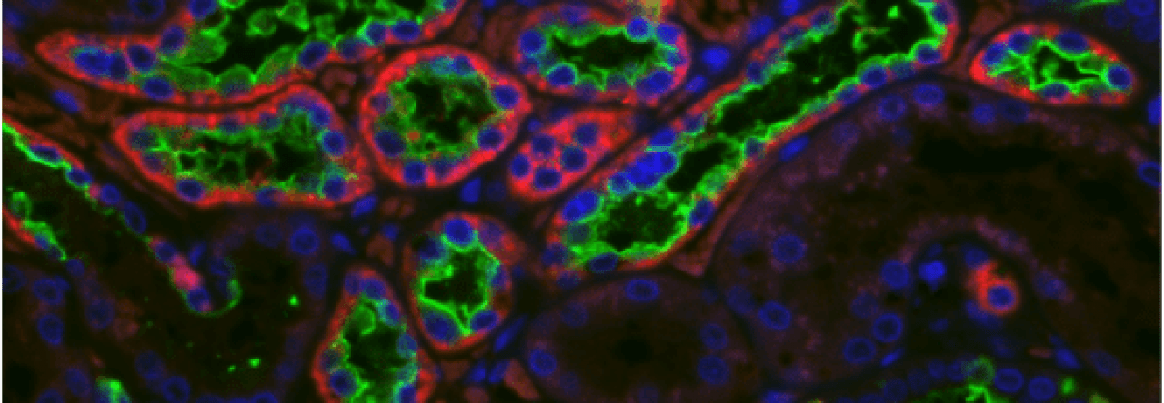
Since viruses are so small, viruses and microscopy have always shared an intimate relationship. The Atwood Laboratory utilizes the extensive bioimaging capabilities provided by the Leduc Bioimaging Facility at Brown University, in addition to our own systems, to visualize aspects of the polyomavirus lifecycle and disease pathogenesis. Members of our group are proficient in light microscopy, fluorescence microscopy, confocal microscopy, transmission electron microscopy, and scanning electron microscopy.

An SVG-A glial cell infected with JC Polyomavirus. SVG-A glial cells were infected with JCPyV for 72 hours. Actin filaments (pseudocolored red) are visible along with JC VP1 (green). This image was formed from the maximum intensity projection of 22 optical slices (thickness = 0.4µm) at 63x magnification. The horizontal field width is 142µm. Sample preparation: K. Garabian, J. Kaiserman, J. Saskin. Image: J. Kaiserman. Visualized with the Zeiss LSM 710, courtesy of the Leduc Bioimaging Facility at Brown University.

JC Polyomavirus virions in purified virus stock. JC Polyomavirus was purified with a cesium chloride gradient and stained using ammonium molybdate. Images were acquired at 153,000x magnification, and the scale bar represents 50nm. Sample preparation: J Kaiserman. Image: J. Kaiserman. Visualized with the Philips 410 TEM, courtesy of the Leduc Bioimaging Facility at Brown University.

JC Polyomavirus virions in viral lysate. The lysate of cells infected with JC Polyomavirus was stained using ammonium molybdate. Images were acquired at 153,000x magnification, and the scale bar represents 50nm. Arrows indicate virions. Sample preparation: J Kaiserman. Image: J. Kaiserman. Visualized with the Philips 410 TEM, courtesy of the Leduc Bioimaging Facility at Brown University.
