All papers and grants listed below are first/last-authored or single-PI’ed by Jonghwan Lee.
Cerebral Microvascular Imaging
We develop and apply OCT techniques for advanced imaging of microvasculature and microcirculation in the brain of disease model animals. OCT (optical coherence tomography) enables label-free, microscopic, three-dimensional imaging of various vascular and cellular dynamics, including blood flow, cellular viability, and neuronal activity. We are specifically interested in applying our techniques to a study on Alzheimer’s disease.
- National Institute of Biomedical Imaging and Bioengineering, K99/R00 ($0.9M, 2013-2018)
- Rhode Island Foundation Medical Research Fund
- Brown University Research Seed Award
- National Institute on Aging, R01 ($1.8M, 2020-2025)
- Multiple-capillary measurement of RBC speed, flux, and density with OCT (2013)
- Statistical intensity variation analysis for rapid volumetric imaging of capillary network flux (2014)
- Early capillary flux homogenization in response to neural activation (2016)
- OCT imaging of capillary reperfusion after ischemic stroke (2016)
- Doppler OCT clutter rejection using variance minimization and offset extrapolation (2018)
- Deep learning toolbox for enhancement, segmentation, and graphing of OCT angiograms (2020)
- Visible-spectrum line-field OCT for high-throughput imaging (2021)
- Deep learning for accurate RBC flux measurement in capillary vessels (2022)
- Viewpoint: Why we need longitudinal study on vascular factors of Alzheimer’s disease (2022)
- Micrometer-resolution line-field OCT (2022)
- Digital micromirror device-based multispectral microscopy (2022)
- Time stretching-based ultrafast holographic microscopy (2022)
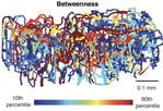
- Longitudinal tracking of pial and penetrating vessel properties in 3xTg mice (2023)
- Longitudinal tracking of all-scale, 25 vascular properties in 3xTg mice (2023)
|
|
 Nat Commun |
 Cover |
Functional Precision Medicine
We develop optical techniques for label-free, multi-metric, 3D cell viability imaging of tissue spheroids toward functional precision cancer medicine.
- Brown University Salomon Faculty Research Award
- Brown University Grant Resubmission Award
- National Cancer Institute, R01 ($1.7M, 2021-2026)
- Dynamic light scattering optical coherence tomography (DLS-OCT) (2012)
- Quantitative imaging of cerebral blood flow velocity and intracellular motility using DLS-OCT (2013)
- Confocal profile and curved focal plane for OCT mapping of the attenuation coefficient (2018)
- Standard-unit measurement of cellular viability using DLS-OCT (2018)
100+ cited |
Photonic Neural Interface
We develop, optimize, and validate novel neural interfaces based on photonics.
- National Eye Institute, R01 ($2.1M, 2019-2024)
- Measurement of fast optical signal of neural activity in brain tissue and its theoretical origin (2010)
- Multiphysics neuron model for cellular volume dynamics (2011)
- Motion correction for phase-resolved dynamic OCT imaging of rodent cerebral cortex (2011)
- Theoretical study on gold nanorod-enhanced near-infrared neural stimulation (2018)
100+ cited |