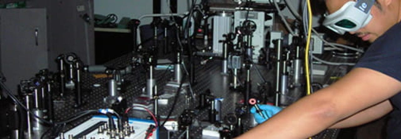
In the Probe Lab we look at many different facets of biophotonics, from Pulse Oximetry, Mueller matrix and Second Harmonic Generation Imaging
Pulse Oximetry
Pulse oximetry is a noninvasive measurement of oxygen saturation level in the blood. Typically it tells us how well a person’s lungs are working at delivering oxygen to the extremities of the body.
Our research is centered around enhancing the precision of pulse oximeters, devices crucial for estimating oxygen saturation levels in the blood. A critical challenge we’ve identified is the impact of skin tone on these measurements, especially for individuals with darker skin, leading to potential overestimations of oxygen levels. This disparity can result in unequal access to vital healthcare, such as black patients facing hurdles in receiving appropriate COVID-19 treatments.
Some of the things we are developing include:
A revolutionary pulse oximeter that integrates skin tone variations into its readings, exploits the electric field properties of light, and precise quantitative assessments of skin tone to provide an accurate reading of the oxygen saturation levels in the blood.
Synthetic tissue platforms for benchmarking optical biomarker measurement technologies. The platform allows for the simulation of various characteristics, such as vasculature and skin melanin content, the latter of which will help close the racial disparity in medical devices technologies such as pulse oximetry.
In tandem we are developing comprehensive simulations to test these optical systems.
Mueller Matrix
The sensitivity of polarized light to certain structures and components makes the Mueller matrix polarimetry a powerful tool for the non-invasive study of biological tissues. The Mueller matrix is mostly used in polarimetry measurements as a linear description of the transformation of the polarized light through the sample, but the interpretation of each element is not straightforward. Although several methods like Mueller matrix polar decomposition and Mueller matrix transformation enable the extraction of several meaningful polarimetric parameters related to three major effects, which are diattenuation, depolarization, and retardance, there is a critical need for quantitative correlation of these parameters with structural and compositional changes. This calls for replicative systems that could act as biological proxies and mimic the characteristic trends in typical biological observations. Among various types of tissue phantoms, we employed the fibroblast-derived tissue construct to explore how the self-assembly process impacts the polarimetric parameters derived from the Mueller matrix. The transforming growth factor beta (TGF-β) treatment enhanced collagen crosslinking and consequently increased the birefringence of the samples. Understanding the correlation between structural changes in collagen-rich tissues and polarimetric parameters could further serve as a quality control in tissue engineering.
Second Harmonic Generation Imaging
Second-harmonic generation (SHG) imaging provides a platform for label-free and non-destructive assessment of specimens, with specificity to non-centrosymmetric materials, such as collagen in tissues. Conventionally, the second harmonic (SH) is generated inside the focal volume of a tightly focused pulsed laser beam, which is then scanned across the sample to build the image, pixel-by-pixel. We have have proven experimentally that it is possible to overcome the speckle noise associated with SHG to render accurate 3D images of collagen.
