Brown University Electron Microprobe Facility
The JEOL JXA-iSP100 Electron Microprobe at Brown University
The JEOL iSP100 electron microprobe is one of the primary and fundamental research instruments in the Department of Earth, Environmental and Planetary Sciences. It is a non-destructive, analytical instrument that is designed to both image the surface of a solid, non-organic sample, such as a rock, mineral or piece of ceramic and determine the chemical composition of small volumes on that sample surface. In 2023-24, through grants from the National Science Foundation (NSF), the National Aeronautics and Space Administration (NASA), and some funding from Brown University, the new microprobe was purchased to replace the aging 22-year-old Cameca SX-100 microprobe, and was installed in September 2024.
The Brown University Electron Microprobe Facility is located in the GeoChem building, room 027.


Installed Options
The JEOL iSP100 microprobe is fully equipped with five wavelength dispersive spectrometers (WDS), a Bruker energy dispersive spectrometer (EDS), and a cathodoluminescence (CL) detector. The WDS spectrometers each contain two diffracting crystals and presently include TAPL, TAP, LPET(3), LLIF(3), LDE1L and LDE6L. The last two crystals are used to measure the light elements (boron to sodium). The absolute detection limits for the instrument are approximately 20-200 ppm depending on the element. The Bruker EDS spectrometer is capable of detecting all elements above beryllium (Be). The microprobe can accommodate a variety of sample types including one inch round slides or polished mounts, rectangular slides, or smaller hand samples that have been flat polished.
Calculating a Composition
To calculate a composition on the microprobe, an electron beam is generated and interacts with bound electrons in the atoms of a flat, polished sample. Electrons from the innermost shells of the bound atoms are scattered, producing vacancies. These vacancies are unstable and are typically quickly filled by electrons from higher electron shells in an atomic cascade. The energy produced as a result of this cascade is emitted from the atom in the form of x-rays. The x-ray wavelengths produced are characteristic of the element(s) present in the sample and the x-ray intensities can be read and measured by the wavelength dispersive spectrometers on the microprobe. By comparing the sample element intensity data to intensity data from mineral standards that have known compositions, the composition of each analysis spot can be calculated.

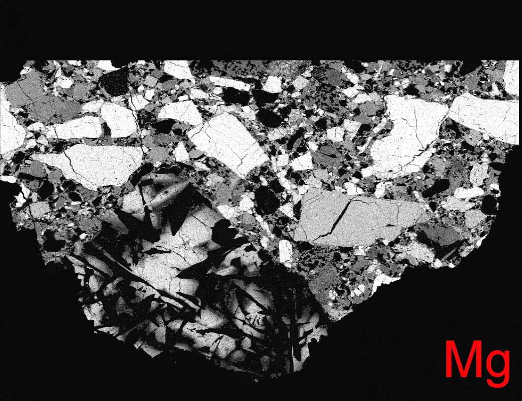
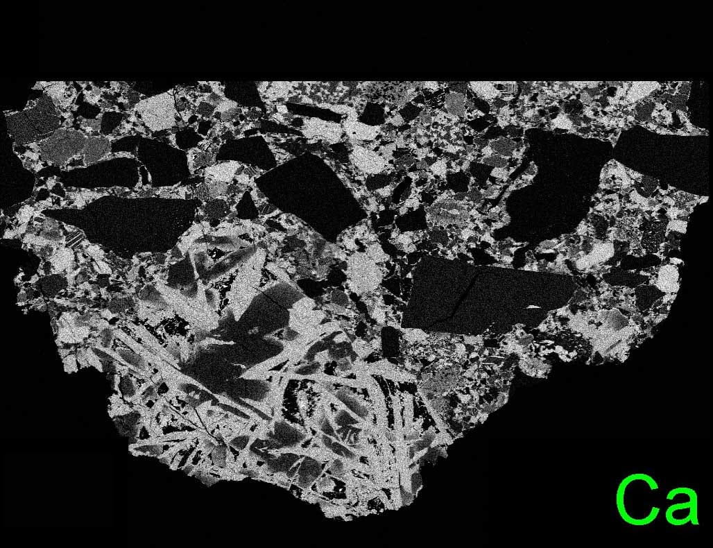
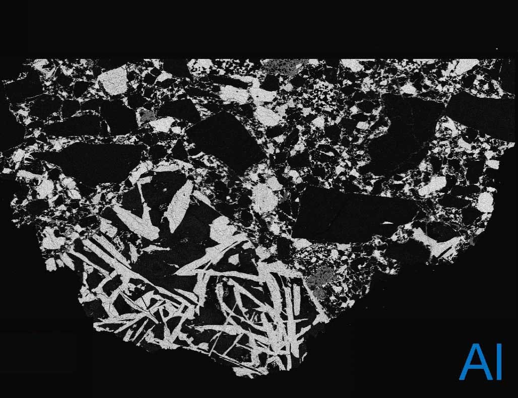
Element X-ray and Phase Mapping
Element x-ray maps can easily be acquired with the microprobe. Using 512×512 pixel maps, up to five element maps, plus a backscattered electron (BSE) image, can be obtained simultaneously. Maximum resolution on any one map is one micron per pixel. Utilizing the stage coordinate system, maps of entire thin sections can be generated. Individual element maps can subsequently be stitched together using free software, such ImageJ. Finally, red-green-blue (RGB) phase maps can be generated by overlaying different element maps to form mineral phase maps. An example is shown below.

Electron Microprobe Sample Holders
A variety of holders are available to accommodate nearly any type of sample. The holders we have at Brown are shown here. Some of the holders were purchased with the instrument, while others were made by our department machinist.

This holder can accommodate ten, one inch round thin sections or polished sections.

A sample holder used for thick, one inch round polished sections or one inch sub-holders, used to hold smaller samples.

A holder for four, rectangular, geologic thin sections and two, one inch round thin sections.

A holder that is primarily for larger samples that are flat on both the top and bottom surfaces. The height of the sample can be adjusted.

This is essentially a tub holder, that when used with a crumpled aluminum foil, can hold many, irregularly shaped samples.

This is a holder from the previous microprobe that I adapted for the new JEOL microprobe. This is primarily for holding four rectangular thin sections, but can accommodate double thick slides and any samples that are under 4 mm thick.

This is a holder that also accommodates larger sample slabs. You just have to make sure you get the height adjusted correctly. If not, you can damage the sample and/or the microprobe.

A sub-holder for use with samples smaller than 0.75 inches in diameter. This sub-holder slips into some of the larger holders.