Brown University Electron Microprobe Facility
The Cameca SX-100 microprobe at Brown University
The Cameca SX-100 electron microprobe is one of the primary and fundamental research instruments in the Department of Earth, Environmental and Planetary Sciences.
In 2014, through an internal research grant from Brown University, the microprobe was upgraded with a new computer running Windows 7, version 5 of Cameca’s Peaksight software, an improved vacuum system, a new electron gun that houses a LaB6 filament, and a joystick.
The Brown University Electron Microprobe Facility is located in the GeoChem building, room 027.


Spectrometers
The Cameca SX-100 electron microprobe is one of the primary and fundamental research instruments in the Department of Earth, Environmental and Planetary Sciences.
In 2014, through an internal research grant from Brown University, the microprobe was upgraded with a new computer running Windows 7, version 5 of Cameca’s Peaksight software, an improved vacuum system, a new electron gun that houses a LaB6 filament, and a joystick.
Installed Options
Capable of analyzing most solid, non-organic materials, it is fully equipped with 5 wavelength dispersive spectrometers (WDS), a Rontec energy dispersive spectrometer (EDS), a secondary electron (SE) detector and a monochromatic cathodoluminescence (CL) detector.
A software option called beam tracking is also installed on this microprobe. This allows the instrument to deflect the electron beam slightly, when necessary, and guarantee that stored stage positions be precisely analyzed within a micron of their original setup position, even hours to days later, despite minor stage drift.
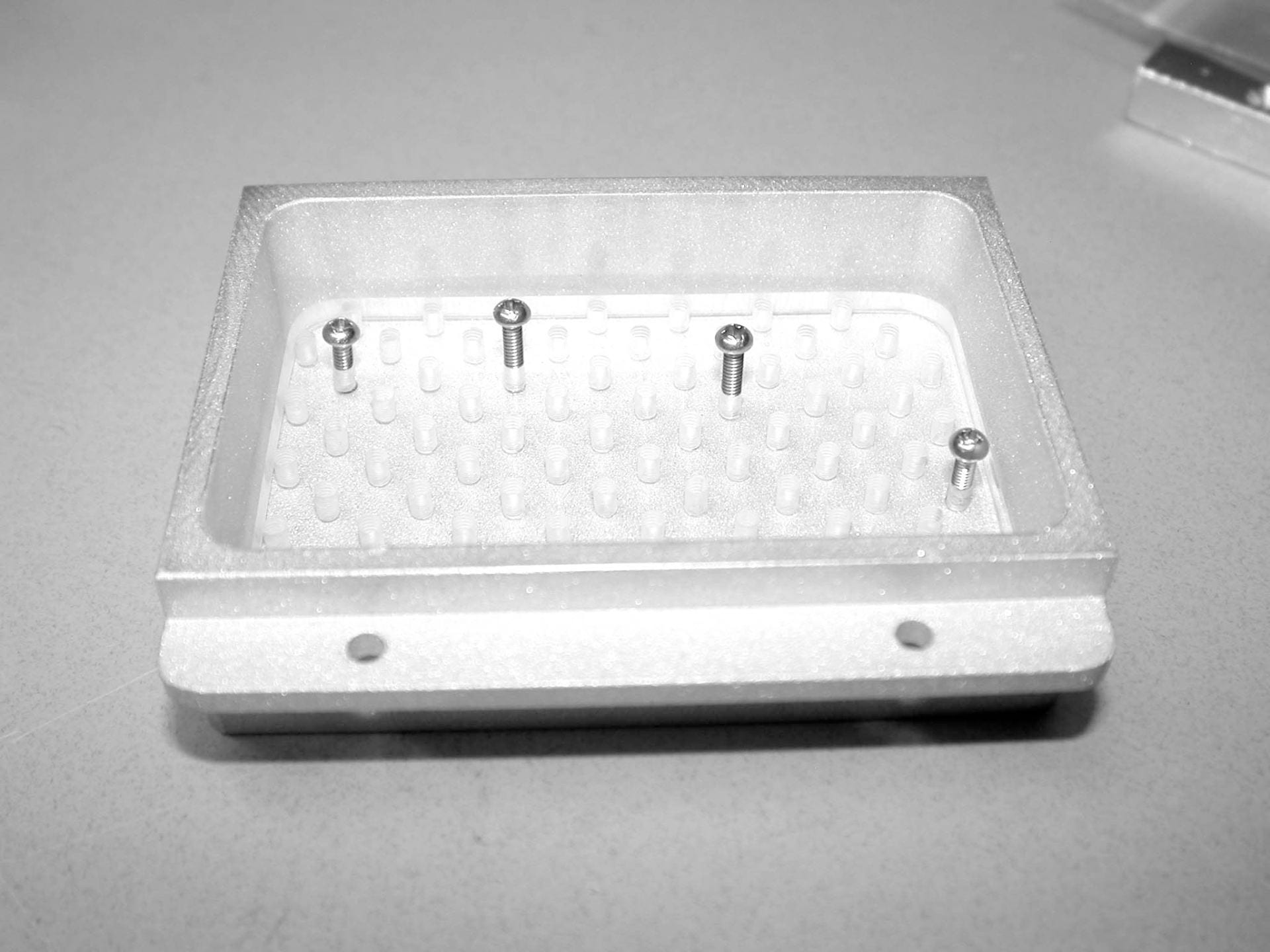
The microprobe can accommodate a variety of sample types including 6 one inch round slides or mounts, 4 rectangular slides or short mounts, or simple hand samples that have been flat polished.

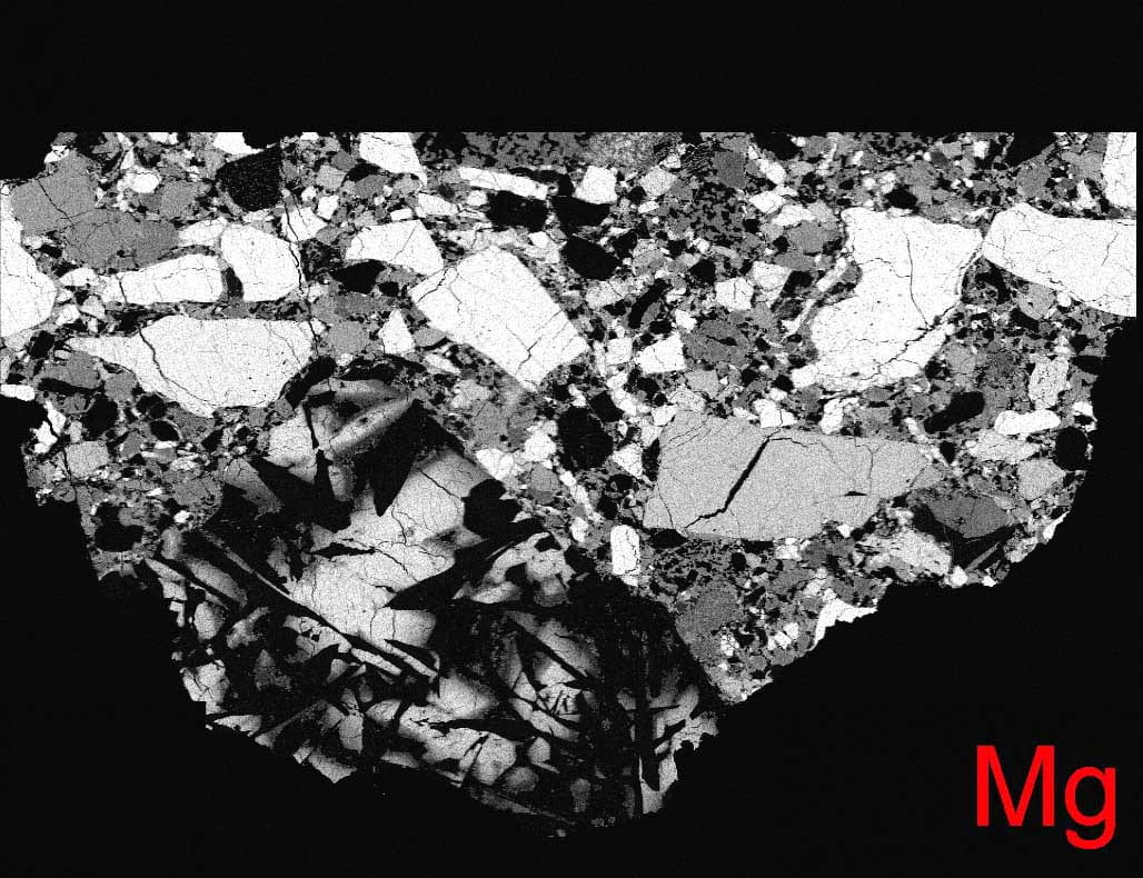
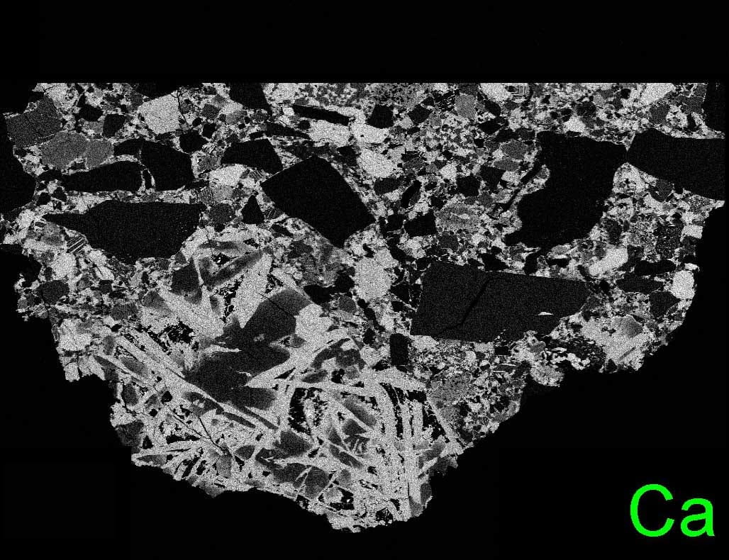
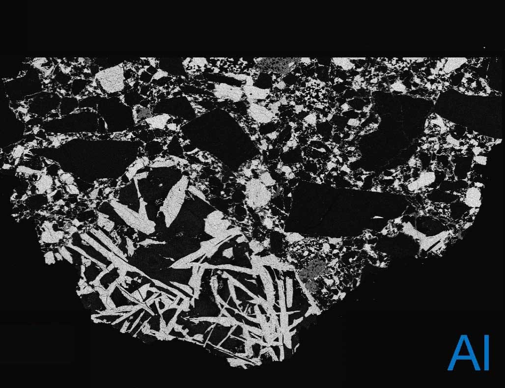
Element X-ray and Phase Mapping
Element x-ray maps can easily be acquired with the microprobe. Using 512×512 pixel maps, up to nine element maps (5 elements on WDS, 4 on EDS), plus a backscattered electron (BSE) image, can be obtained simultaneously. Maximum resolution on any one map is one micron per pixel. Utilizing the stage coordinate system, maps of entire thin sections can be generated. Individual element maps can subsequently be stitched together using ImageJ software. Finally, red-green-blue (RGB) phase maps can be generated by overlaying different element maps. An example is below.
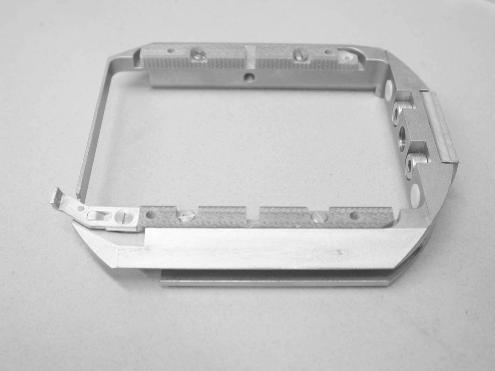
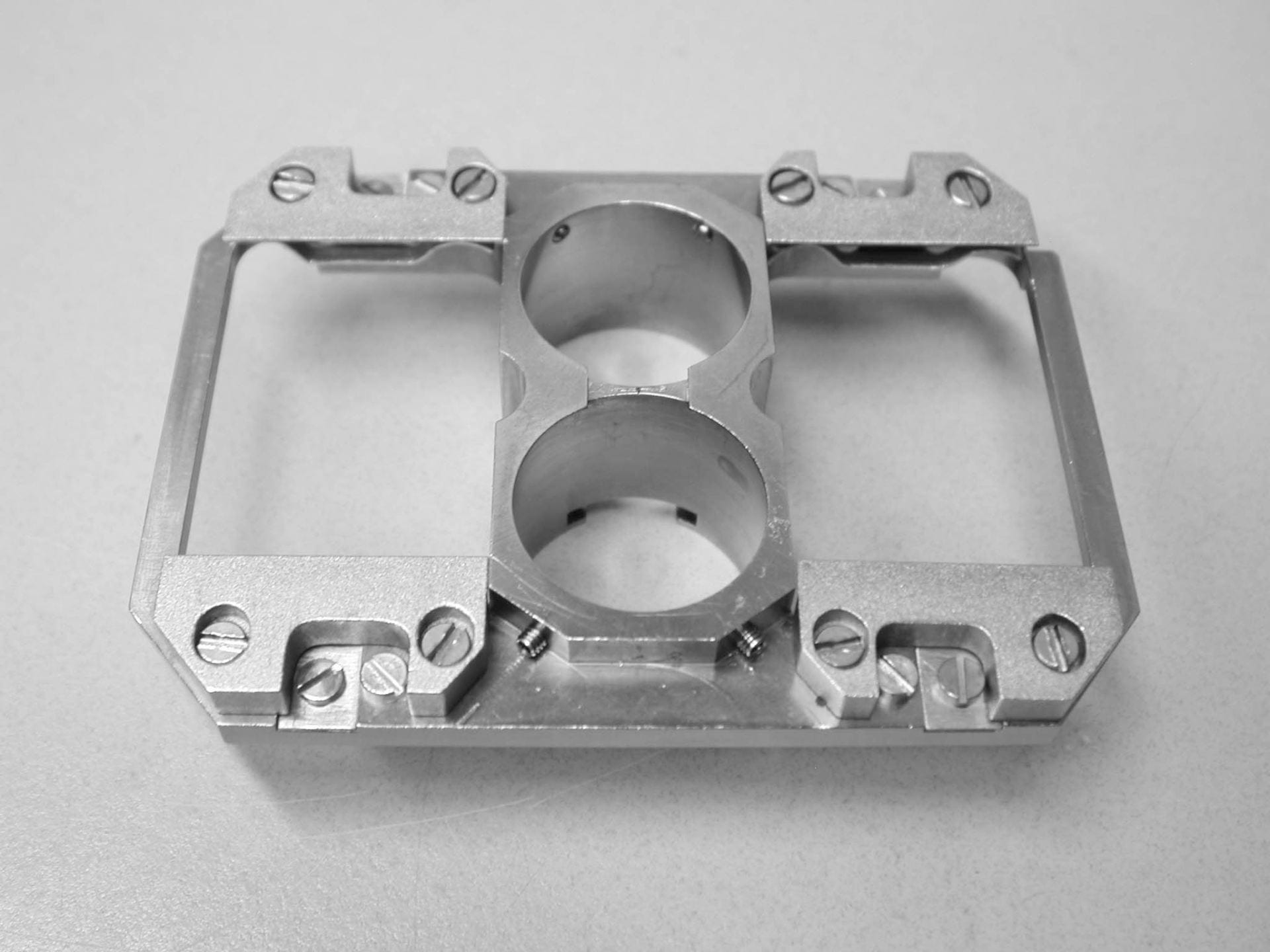
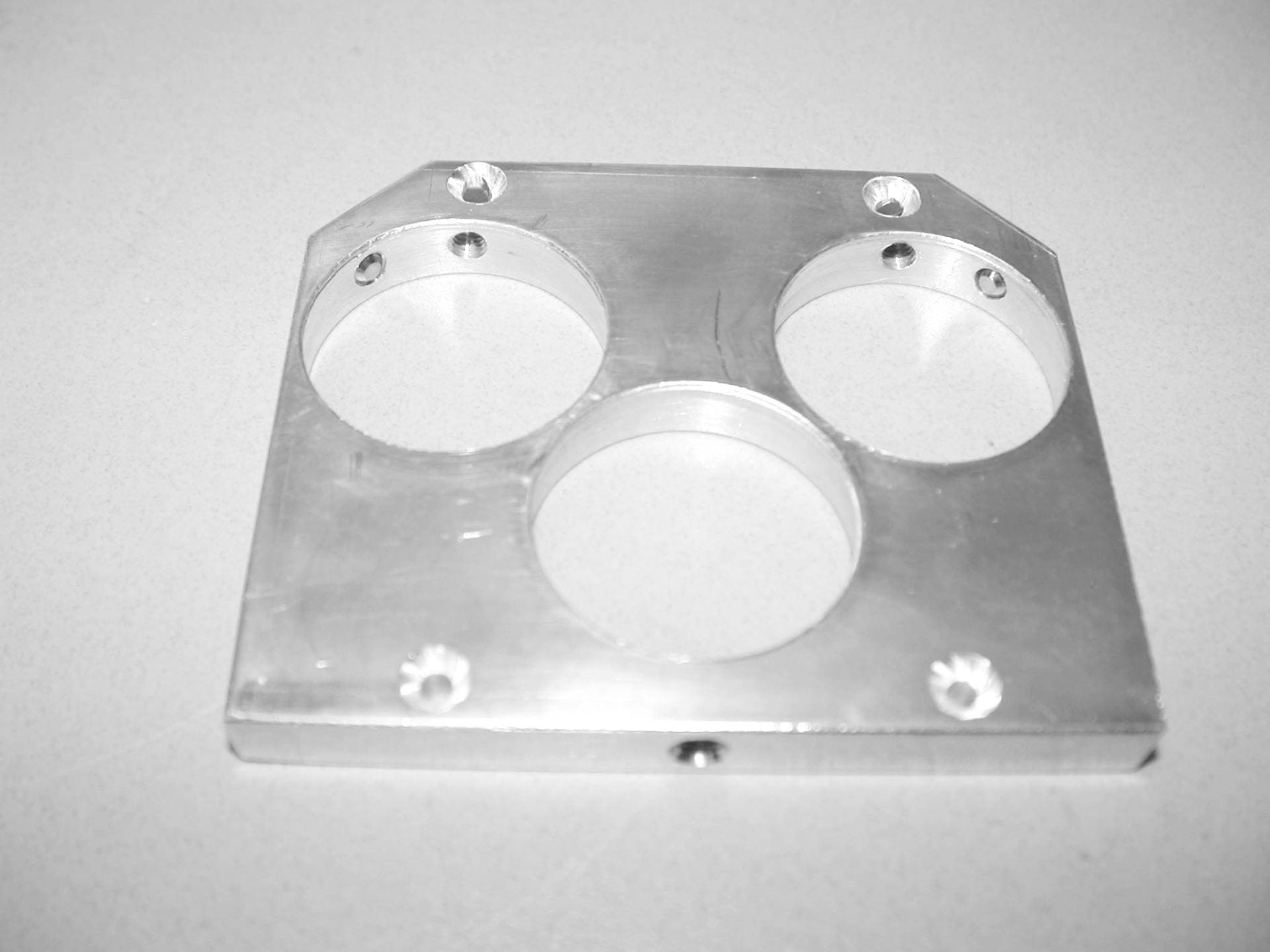
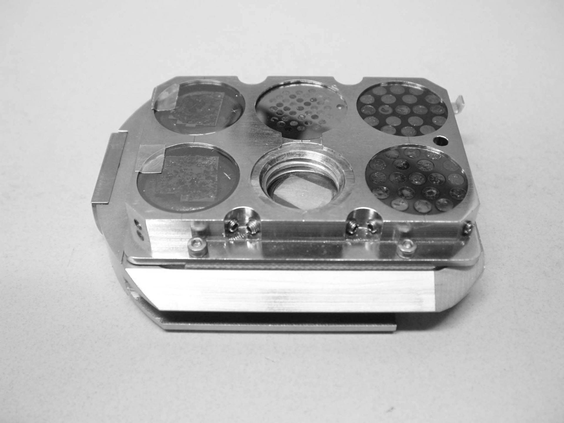
A variety of holders can be accommodated. The one’s we have at Brown are shown here. Some of the holders we purchased with the instrument, some were made by our machinist.

Electron Microprobe Sample Holders
The Cameca SX-100 electron microprobe has a two part shuttle system that is used to hold and insert samples into the instrument. The shuttle consists of a lower rail and an upper holder.
A variety of holders can be accommodated. The one’s we have at Brown are shown here. Some of the holders we purchased with the instrument, some were made by our machinist.