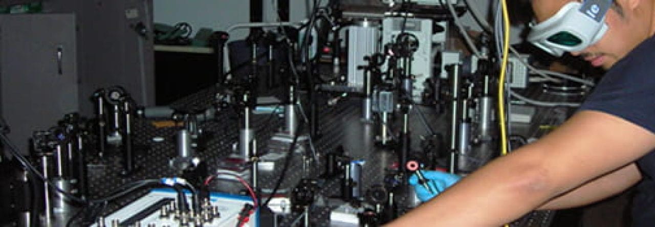
For several centuries, optical microscopy has been defined by the variety of methods by which objects too small to see with the naked eye can be imaged. However, recent progress in the biomedical sciences has called for optical imaging techniques that improve on spatial and temporal resolution, contrast, and specificity to features of interest—ideally without damaging the specimen under study. An equally important characteristic of 21st-century imaging is that the new modalities facilitate quantitative assessment through the employment of straightforward image processing techniques.The rule of thumb being that there is a tradeoff between complexity of the image processing technique and the discernible information in the raw image. At PROBE lab our attempt at this problem has led to Fourier transform-second-harmonic generation (FT-SHG) microscopy. We effectively combine the nonlinear imaging technique of SHG with spatial harmonic analysis (using Fourier transforms) to image fibrillar collagen-based tissues. Similar to other nonlinear microscopy techniques, SHG permits sub-micron 3D spatial resolution at optical wavelengths, reduced phototoxicity, and high contrast. Unlike two-photon fluorescence microscopy, SHG permits direct imaging of structural proteins, which exhibit noncentrosymmetry, such as fibrillar collagen, myosin, and microtubules without the need for staining. We have shown that spatial harmonic analysis is well-suited for such a technique. In particular, the combination of the two, FT-SHG has been applied to quantifying the organization of collagen fibers (e.g., fiber orientation and packing density) in tissues such as the ear, tendon, eye, and bone [Opt. Exp. 17, 14534 (2009)].
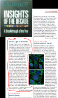 We have also shown that, with respect to spatial organization, the information content between the forward-scattered and backward-scattered SHG signal is very similar, making the technique attractive for potential diagnostic applications [Opt. Lett., 34, 3779 (2009)]. In addition, a recent demonstration of FT-SHG has successfully differentiated between healthy and injured tendons (this work was featured in the Editor’s Choice biophysics segment of Science) [Opt. Exp. 18, 24983 (2010)]. Another interesting feature of SHG microscopy that differs from its two-photon fluorescence counterpart is that the process conserves energy, and thus phase information between the applied input field and the resultant second-harmonic signal is preserved. This opens up possibilities for using the optical polarization to extract quantitative information, below the diffraction limit, encoded in the second-harmonic scattering coefficient. The PROBE lab is currently exploring combining this approach with FT-SHG in order to quantify both the structure and content of tissues. Ultimately, we believe that such a tool will be useful in assessing changes in collagen fiber organization and/or content resulting from either damage or disease.
Finally, in collaboration with researchers at the University of Minnesota, and sponsored by the National Academies Keck Futures Initiative (NAKFI), we are working to introduce a new paradigm for intelligent interactive imaging that combines SHG microscopy with 3-D user interface and data visualization technology in order to leverage the best of both human and machine intelligence for smarter imaging.
![SHG image of injured horse tendon [adapted from Opt. Exp. 18, 24983 (2010)]](http://publish.illinois.edu/mechse-probe/files/2014/09/shg_tendon_image.20110923.4e7ccfbc4f6894.79283194.png)
SHG image of injured horse tendon [adapted from Opt. Exp. 18, 24983 (2010)]